XB-IMG-128966
Xenbase Image ID: 128966
|
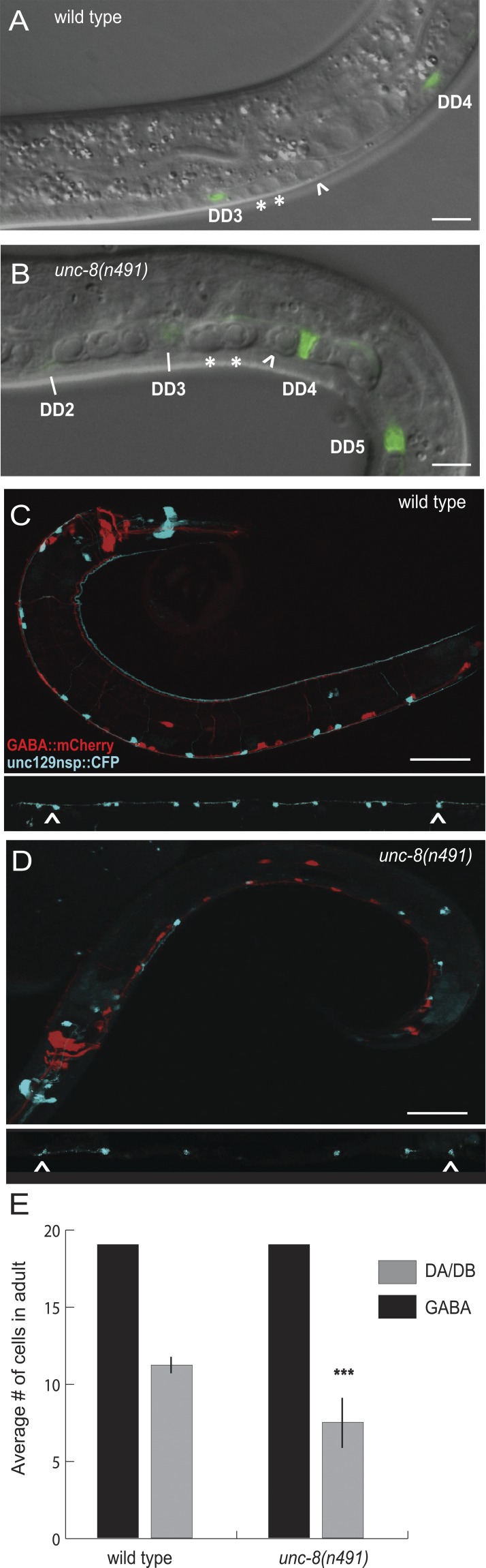
Figure 1. Selective degeneration of ventral cord cholinergic motor neurons in unc-8(n491) animals. DIC images of L1 larval ventral nerve cords of (A) wild-type versus (B) unc-8(n491) animals. Cholinergic DA and DB motor neurons (asterisks) and P cell precursor (arrowhead) appear swollen in unc-8(n491) versus wild type. DD GABA motor neurons are labeled with punc-25::GFP. Bar, 1 µm. (C and D) DD/VD GABAergic motor neurons are marked with punc-25::dsRed (red) and DA/DB neurons are labeled with unc-129nsp::CFP (blue) in (C) wild-type and (D) unc-8(n491) young adult animals. Insets show linearized view of DA/DB motor neurons. Bars, 25 µm. (E) GABAergic and DA/DB motor neurons were counted in the ventral cord region between DD1 and DD6 (arrowheads in C and D; see also Fig. S1). unc-8(n491) animals (n = 14) contain the full complement of 19 DD/VD GABAergic motor neurons but are missing a significant fraction (∼36%) of the 11 DA/DB motor neurons from this region of the adult ventral nerve cord (wild type; n = 24). ***, P = 3 × E-11 by t test. Image published in: Wang Y et al. (2013) © 2013 Wang et al. Creative Commons Attribution-NonCommercial-ShareAlike license Larger Image Printer Friendly View |