XB-IMG-117400
Xenbase Image ID: 117400
|
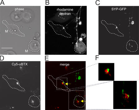
Figure 5. Synaptic sites are revealed by double labeling with SYP-GFP and Cy5-α-BTX. (A) Phase-contrast image of nerve-muscle coculture. M, myocytes. (B) Labeling of axons of a spinal neuron by rhodamine dextran. (C) Labeling of synaptic varicosities by SYP-GFP. Note the discrete SYP puncta located in presynaptic terminals innervating the myocytes. (D) Labeling of postsynaptic AChRs with Cy5-α-BTX. (E) Merge of images in C and D. (F) Enlargement of the areas boxed in E. Note that SYP-GFP (green) and Cy5-α-BTX (red) puncta are juxtaposed against each other at the synapses. Bar, 10 μm. Image published in: Je HS et al. (2006) Copyright © 2006, The Rockefeller University Press. Creative Commons Attribution-NonCommercial-ShareAlike license Larger Image Printer Friendly View |