XB-IMG-171101
Xenbase Image ID: 171101

|
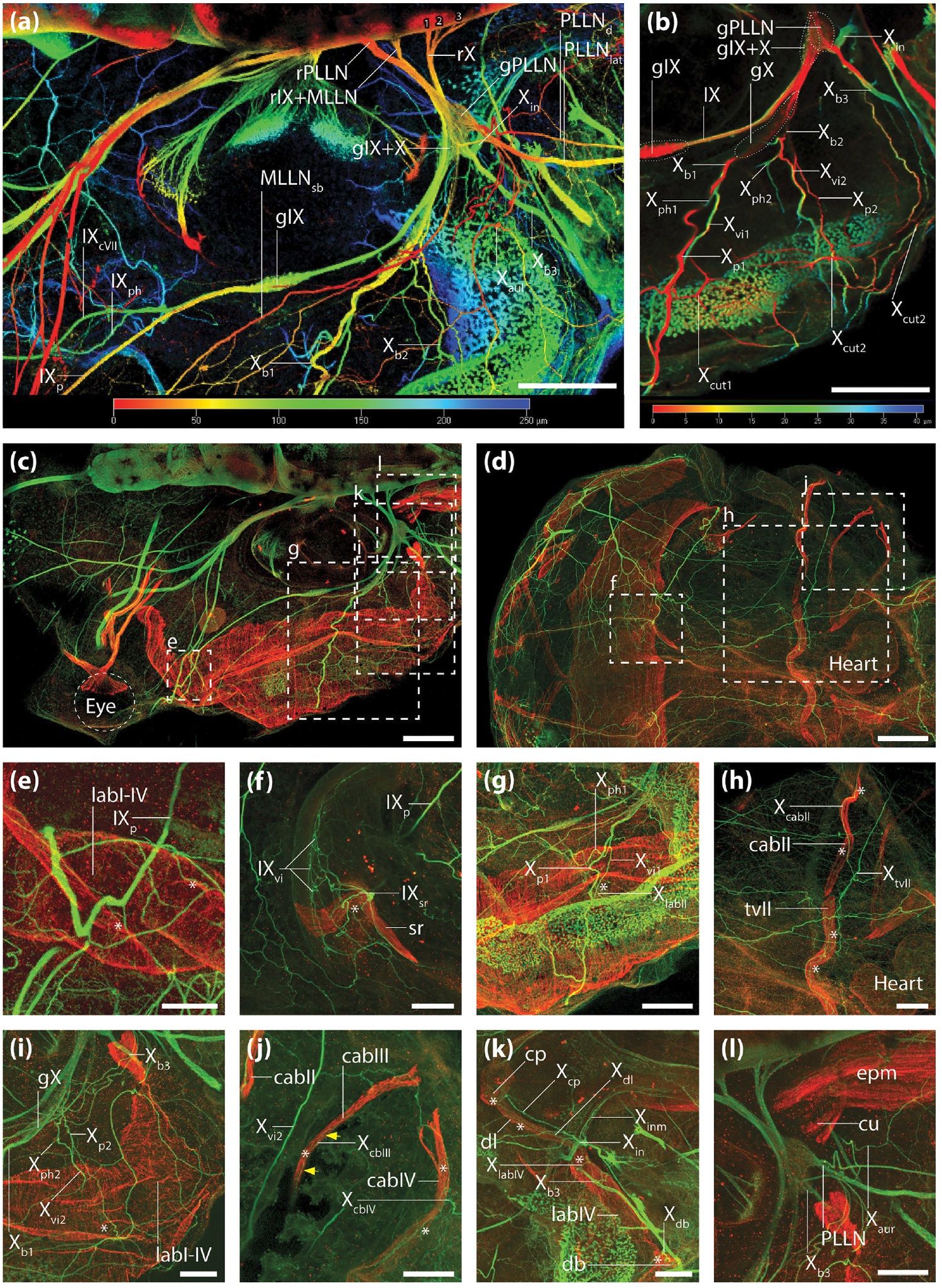
FIGURE 8 The glossopharyngeal and vagal nerves. Whole mount antibody staining of the nerves (anti-acetylated alpha-tubulin) and
muscles (anti-desmin) of a NF stage 47/48 X. laevis tadpole. (a and b) 3D depth coding views of the nerves. (a) dorsal view of the region of
the otic capsule. (b) dorsal view of the distal branchial region. (câl) maximum intensity projections of the same digital stack. Nerves are
shown in green, muscles are shown in red. (c) dorsal overview of head and branchial region. (d) ventral overview of the head and branchial
region. (eâl) magnifications of the areas marked by the strippled rectangles in (c) and (d). The white asterisks indicate the innervation sites
of the branchial muscles by rami/ramuli of the glossopharyngeal (IX) and vagal nerves (X). The yellow arrows indicate the route of the
ramulus constrictor arcus branchiarium III of the X. (l) dorsal view of the cucullaris muscle. The white scale bar in (c) and (d) is 200 mm, in (e)
to (l)50 mm [Color figure can be viewed at wileyonlinelibrary.com] Image published in: Naumann B and Olsson L (2018) Copyright © 2018. Image reproduced with permission of the Publisher, John Wiley & Sons.
Image source: Published Larger Image Printer Friendly View |
