XB-IMG-121071
Xenbase Image ID: 121071
|
|
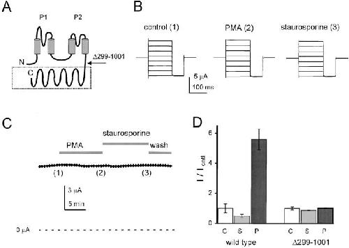
Figure 4. Truncated (KCNKΔ299-1001) channels are not subject to regulation. Macroscopic KCNKØ currents were measured as in Fig. 1. (A) Predicted membrane topology of KCNKØ indicating two P loop domains (P1 and P2), four transmembrane segments and the ∼700-residue segment that is deleted in KCNKΔ299-1001 subunits (boxed). (B) Raw current traces for an oocyte expressing KCNKΔ299-1001 channels under various conditions by the protocol in Fig. 1 B: (1) control solution; (2) after 10-min perfusion with PMA; and (3) after 10-min perfusion with staurosporine. (C) Currents measured during application of control, PMA, and staurosporine solutions. The oocyte was held at −40 mV and stepped to 25 mV for 250 ms with a 20-s interpulse interval. Data for B was collected at the indicated times. (D) Normalized mean currents measured in groups of 17 oocytes at 25 mV after 20- min incubation in the following: (C) 20 mM potassium solution (open bars); (S) 2 μM staurosporine (gray bars); or (P) 50 nM PMA (black bars). Bars are mean ± SEM for current in experimental compared with control solution. Image published in: Zilberberg N et al. (2000) © 2000 The Rockefeller University Press. Creative Commons Attribution-NonCommercial-ShareAlike license Larger Image Printer Friendly View |