XB-IMG-117890
Xenbase Image ID: 117890
|
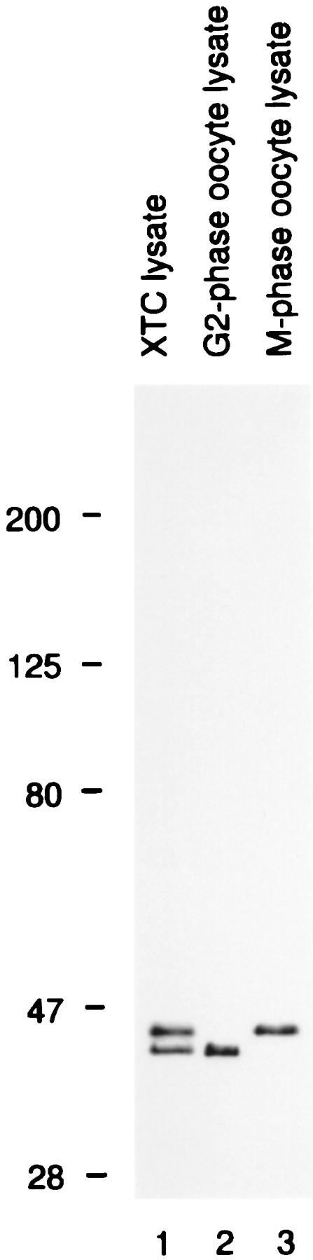
Figure 1. The specificity of antibody X15. Immunoblots of lysates from XTC cells (lane 1), G2-phase oocytes (lane 2), and M-phase oocytes (lane 3) were probed with X15. XTC cells exhibited two bands: a lower band that comigrated with nonphosphorylated Erk2, and an upper band that comigrated with both Erk1 and phosphorylated Erk2. The upper band was identified as Erk1, rather than phosphorylated Erk2, based on its cross-reactivities with other Erk1 and Erk2 antisera, and on the fact that it did not shift to a lower apparent molecular weight when lysates were treated with XCL100 (not shown). Image published in: Wang XM et al. (1997) Image reproduced on Xenbase with permission of the publisher and the copyright holder. Creative Commons Attribution-NonCommercial-ShareAlike license Larger Image Printer Friendly View |