XB-IMG-154657
Xenbase Image ID: 154657
|
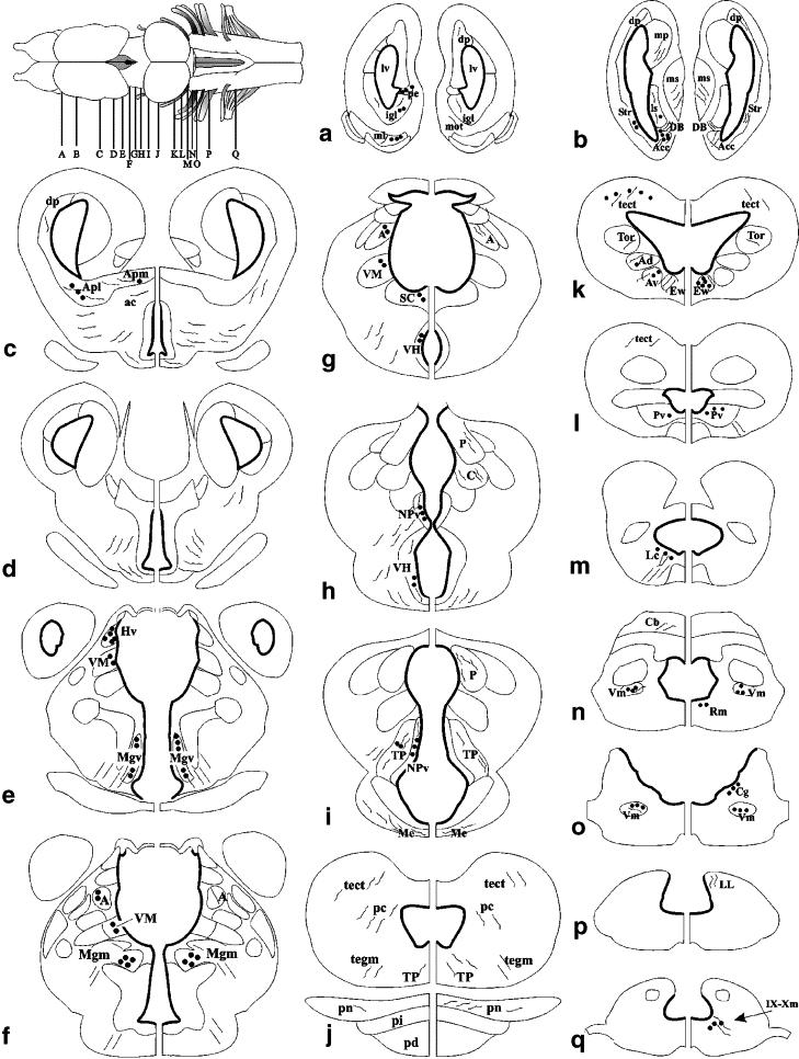
Fig. 1.
Top left: schematic dorsal view of the brain of X. laevis, with letters aâq indicating the levels of transverse sections (aâq) used in the study of CRF (on the left) and UCN1 (on the right) distribution. Immunoreactive cell bodies are indicated by black dots and immunoreactive main axon tracts by lines. A, anterior thalamic nucleus; ac, anterior commissure; Acc, nucleus accumbens; Ad, anterodorsal tegmental nucleus; Apl, amygdala, pars lateralis; Apm, amygdala, pars medialis; Av, anteroventral tegmental nucleus; Cb, cerebellum; DB, diagonal band of Broca; dp, dorsal pallium; EW, EdingerâWestphal nucleus; Hv, nucleus habenularis ventralis; igl, internal granule cells of the olfactory bulb; Lc, locus coeruleus; ls, lateral septum; lv, lateral ventricle; me, median eminence of the hypothalamus; Mgm, medial part of the magnocellular nucleus; Mgv, ventral part of the magnocellular nucleus; ml, mitral cell layer of the olfactory bulb; mot, medial olfactory tract; mp, medial pallium; ms, medial septum; NPv, nucleus of the paraventricular organ; nIX, nucleus motorius of the glossopharyngeus (glp) nerve; P, posterior thalamic nucleus; pc, posterior commissure; pd, pituitary gland, pars distalis; pe, post-olfactory eminence; pi, pituitary gland, pars intermedia; pn, pituitary gland, pars nervosa; Rm, nucleus reticularis medius; SC, suprachiasmatic nucleus; Str, striatum; tect, mesencephalic tectum; tegm, mesencephalic tegmentum; Tor, torus semicircularis; TP, posterior tubercle; VH, ventral hypothalamic nucleus; VM, ventromedial thalamic nucleus; Vm, nucleus motorius nervi trigemini; Xm, nucleus motorius nervi vagi. Image published in: Calle M et al. (2005) Copyright © 2005. Image reproduced with permission of the Publisher. Larger Image Printer Friendly View |