XB-IMG-117578
Xenbase Image ID: 117578
|
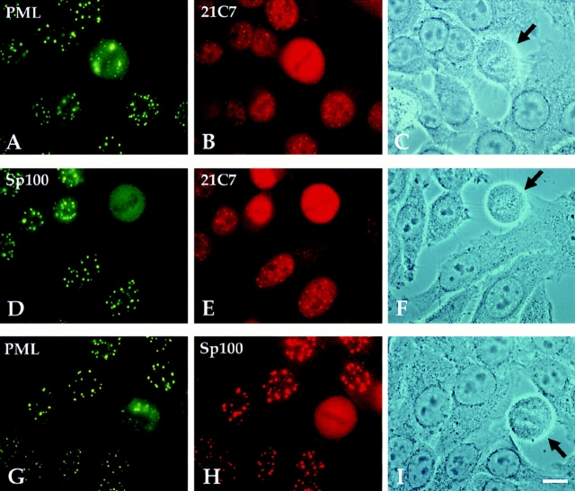
Figure 6. Immunofluorescence staining of methanol/ acetone-fixed, IFN-treated HeLa S3 cells. Cells were either double-labeled with polyclonal rat anti-PML (green) and mAb 21C7 (red) (A–C), with polyclonal rabbit anti-Sp100 antibodies (green) and mAb 21C7 (red) (D to F), or with anti-PML Abs and anti-Sp100 Abs (G–I). In a mitotic cell, PML is aggregated at the periphery of the cell (A and G, arrow), whereas Sp100 and PIC1/ SUMO-1 are diffusely distributed (B, D, E, and H), indicating that PIC1/SUMO-1 and Sp100 dissociate from PML in mitosis. Bar, 10 μm. Image published in: Sternsdorf T et al. (1997) Image reproduced on Xenbase with permission of the publisher and the copyright holder. Creative Commons Attribution-NonCommercial-ShareAlike license Larger Image Printer Friendly View |