XB-IMG-122720
Xenbase Image ID: 122720

|
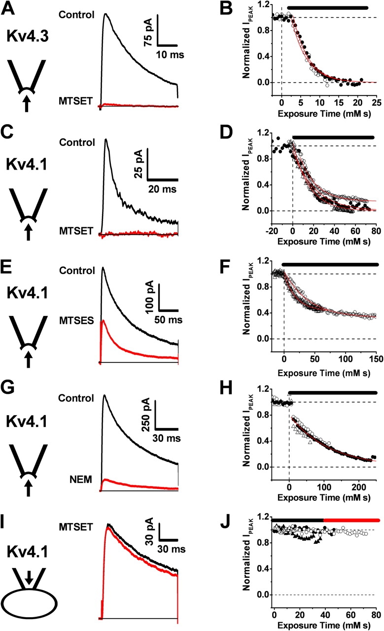
Figure 1. Time-dependent inhibition of Kv4 channels by thiol-specific reagents applied to the intracellular side of the membrane. (A–H) Currents were evoked by a step depolarization from −100 mV to +50 mV at 3-s intervals. The intracellular side of inside-out macropatches from Xenopus oocytes was exposed to the thiol-specific reagents (200–600 μM MTSET, 200 μM MTSES, 2 mM NEM). The current traces are averages of ∼7–20 individual responses recorded before the reagent application (black) and after the inhibition approached steady-state (red). Reagent exposure in the corresponding inhibition time courses (B, D, F, and H) is indicated by a black horizontal bar. Each symbol type represents a separate macropatch (B, n = 2; D, n = 3; F, n = 2; H, n = 3; J, n = 4). Red lines in these graphs are the best fits assuming an exponential decay. The mean values of the derived second-order rate constants (1/(τ × [reagent])) were 248, 58, 37, and 8 M−1s−1, for B, D, F, and H, respectively. (I and J) When MTSET (600 μM) was present in the pipette (external solution), the current remained stable. The black and red traces correspond to the average currents obtained during the first and second half of the experiment, respectively (black and red bars). Image published in: Wang G et al. (2005) Copyright © 2005, The Rockefeller University Press. Creative Commons Attribution-NonCommercial-ShareAlike license Larger Image Printer Friendly View |
