XB-IMG-130089
Xenbase Image ID: 130089
|
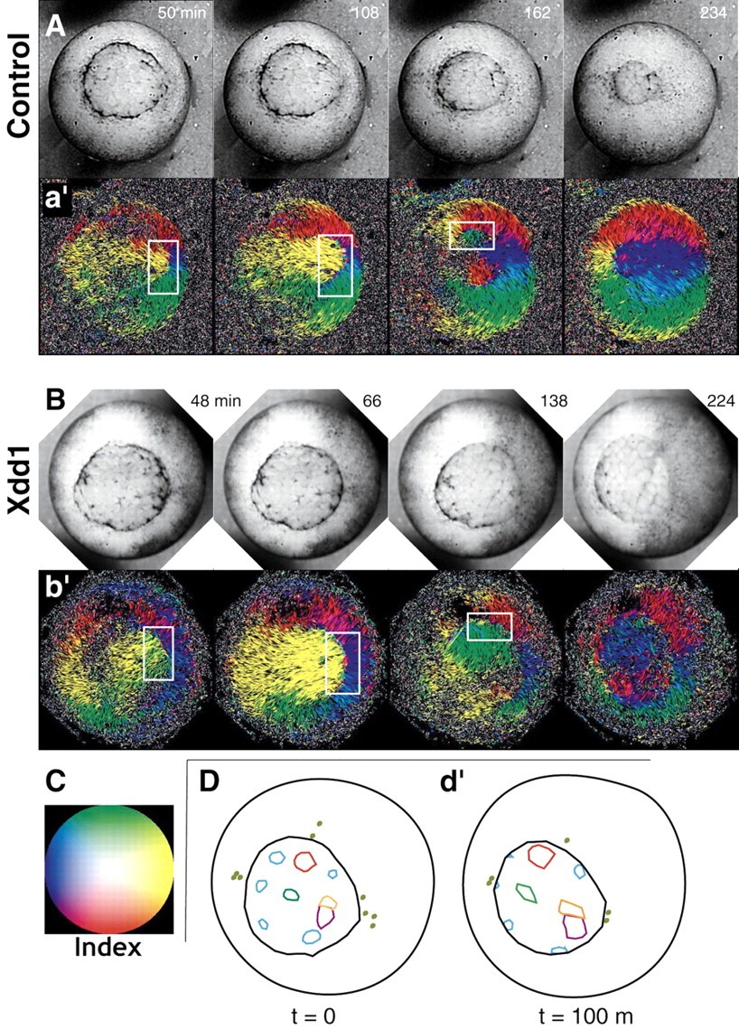
Fig. 5. Marginal zone internalization without blastopore closure in Xdd1-expressing embryos. Optical flow analysis of control (A; see Movie 2 in supplementary material) and Xdd1-injected (B; see Movie 3 in supplementary material) embryos. As indicated in the key in panel C, color denotes the direction of flow in aâ² and bâ² (e.g. yellow lines indicate dorsal movement; blue lines indicate ventral movement). Opposed flow (e.g. yellow meeting blue) represents internalization and is highlighted with white boxes. Cell tracings of Xdd1-injected embryos (D,dâ²) also reveal the internalization of cells in both the marginal zone and yolk plug. Image published in: Ewald AJ et al. (2004) Copyright © 2004. Image reproduced with permission of the Publisher and the copyright holder. This is an Open Access article distributed under the terms of the Creative Commons Attribution License. Larger Image Printer Friendly View |