XB-IMG-130078
Xenbase Image ID: 130078
|
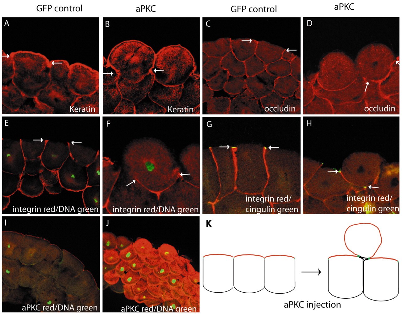
Fig. 3. aPKC is sufficient to promote apical and inhibit basal lateral membrane identity without disrupting tight junctions. (A,B) aPKC overexpression (B) caused expansion of the apical marker keratin compared with GFP control (A). (C-F) aPKC caused reduction in the basolateral markers, occludin (D) and β1-integrin (F) compared with controls (C,E). (G,H) aPKC caused tight junctions (as marked by cingulin) to be maintained but relocated to the new apicobasolateral border. The borders of the markers used in each panel are delineated with arrows. (I,J) aPKC staining in GFP-injected controls (I) and aPKC staining in aPKC-injected (J) embryos. The apicalised cells have inherited overexpressed aPKC. (K) Diagrammatic representation of the result; aPKC causes protruding hyper-apical cells, which still have tight junction markers. Apical, red; basolateral, black; tight junctions, green. Albino embryos were injected with aPKC RNA and stained for antibody markers of cell polarity. Each experiment was carried out three times. Image published in: Chalmers AD et al. (2005) Copyright © 2005. Image reproduced with permission of the Publisher and the copyright holder. This is an Open Access article distributed under the terms of the Creative Commons Attribution License. Larger Image Printer Friendly View |