XB-IMG-175646
Xenbase Image ID: 175646
|
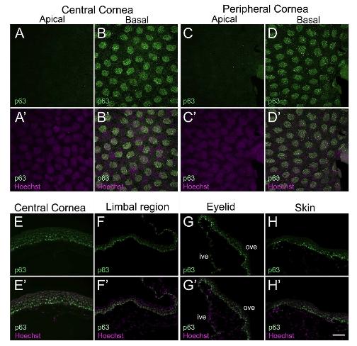
Fig. 2. Confocal and fluorescence light microscopic images showing p63 antibody staining in X. laevis epithelial tissues. (AâD) Larval cornea whole-mounts showing p63 expression (green) is restricted to all basal cells (nuclei) of the central and peripheral cornea, as labeled. (Aâ²-Dâ²) Merged images for A-D with Hoechst labeled nuclei (magenta). (A-Aâ²) Apical cells of central cornea exhibit no p63 expression. (B-Bâ²) Localized p63 labeling is seen in all nuclei of the basal epithelium. (C-Câ²) p63 is not expressed in apical cells of peripheral corneal epithelium. (D-Dâ²) All nuclei of basal cells in the peripheral cornea have uniform expression for p63. (EâH) Localization of p63 (green) in cryosections of adult cornea, eyelid and skin. The apical surface is located towards the top of each image, and the basal surface towards the bottom. (Eâ²-Hâ²) Merged images for E-H with Hoechst labeled nuclei (magenta). (E-Eâ²) p63 labeling is detected in nuclei of basal and intermediate cells of the central cornea, but not in the nuclei of apical cells. The nuclei in the basal epithelium have relatively higher levels of p63 staining than those in the intermediate layer. (F-Fâ²) In the limbal region, p63 label is primarily detected in nuclei of basal epithelial cells along with moderate expression in intermediate layer cells. (G-Gâ²) Ventral eyelid showing p63 staining is localized to the nuclei of cells present mainly in the outer surface of the eyelid, with the cells in basal layer showing higher staining intensity. Some weak expression maybe seen in scattered basal cells within the inner surface of the eyelid. (H-Hâ²) p63 expression is also detected in the surrounding skin epithelium, with nuclei in the basal layer showing higher expression compared to those of the intermediate layer cells. No staining is detected in cells of the outermost, apical layer of the skin. ive, inner ventral eyelid; ove, outer ventral eyelid. Scale bar in Hâ² equals 25â¯Î¼m for A-D, Aâ²-Dâ², and 50â¯Î¼m for E-H, Eâ²-Hâ. (For interpretation of the references to colour in this figure legend, the reader is referred to the Web version of this article.) Image published in: Sonam S et al. (2019) Copyright © 2019. Image reproduced with permission of the Publisher, Elsevier B. V.
Image source: Published Larger Image Printer Friendly View |