XB-IMG-124448
Xenbase Image ID: 124448
|
|
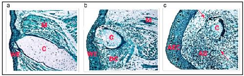
Figure 1. Histology of axolotl hindlimbs. Longitudinal sections of axolotl hindlimbs regenerating from the mid-tibia/fibula, stained with Weigert's iron hematoxylin and light green SF: (a) Sections at 1 day post amputation (dpa). The amputation surface is covered with several layers of wound epidermal (WE) cells, including gland cells. The basal layer of the wound epidermis is in direct contact with underlying tissues. Some cell debris, red blood cells and lymphocytes are present under the wound epithelium. C = cartilage, M = muscle. (b) Sections at 4 dpa. The cartilage (C), muscle (M), and dermal tissue organization is breaking down, releasing cells that dedifferentiate (DC) and migrate toward the wound epithelium (WE). (c) Sections at 7 dpa. Blastema cells have accumulated under a thickened apical epidermal cap (AEC) to form an accumulation blastema (AB). C = cartilage. The arrows indicate the junction between the accumulation blastema and tissues still undergoing dedifferentiation. Magnification = 10 ×. Image published in: Rao N et al. (2009) Copyright ©2009 Rao et al; licensee BioMed Central Ltd. Creative Commons Attribution license Larger Image Printer Friendly View |