XB-IMG-169417
Xenbase Image ID: 169417
|
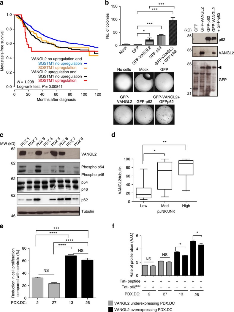
Figure 7. Disruption of the VANGL2–p62/SQSTM1 interaction in breast cancer cells.(a) Kaplan–Meier MFS curves of breast cancer patients according to concomitant VANGL2 and p62/SQSTM1 mRNA expression. The 5-year MFS are 49% (both upregulated; N=27), 64% (both not upregulated; N=833) and 56% (one upregulated, the other not upregulated; N=79 and N=269). (b) Soft agar colony formation of T47D cells overexpressing GFP, GFP–VANGL2 and GFP–p62/SQSTM1 (right). Protein expression was revealed with anti-p62/SQSTM1, anti-VANGL2 and anti-GFP antibodies by western blot analysis (right). In anti-GFP blot, the arrowhead indicates position of co-migrating GFP–VANGL2 and GFP–p62/SQSTM1 and the asterisk pinpoints GFP alone. Error bars represent mean±s.d. (n=3). (c) Protein levels of VANGL2, phosphorylated JNK, JNK (p54/p46), p62/SQSTM1 and tubulin assessed in eight breast cancer PDXs (PDX 1–8) by western blot analysis. (d) VANGL2, JNK, phosphorylated JNK and tubulin signals from 30 PDX protein extracts were quantified. VANGL2/tubulin ratios were plotted against pJNK/JNK ratios, arranged in ascending order into three equally sized groups (low, medium and high). High expression of VANGL2 protein is correlated to high levels of phosphorylated JNK. Box and whisker plots show the median value and interquartile ranges. The Kruskal–Wallis test was used for comparison of the median levels of expression. Statistically significant differences are indicated (*P≤0.05; **P≤0.01). (e) Treatment of the indicated PDX-derived cells (PDX. DC-2, −27, −13 and −26) with a Tat-conjugated JNK inhibitor (Tat-JIP at 10 μM) during 48 h led to greater reduction in cell proliferation of VANGL2high/pJNKhigh than VANGL2low/pJNKlow PDX-derived cells. Comparisons use Tukey's multiple comparisons test. NS, not significant. Data are representative of three independent experiments; *P≤0.05; **P≤0.01; ***P≤0.001; ****P≤0.0001. (f) Treatment of the indicated PDX-derived cells (PDX. DC-2, −27, −13 and −26) with the p62DN peptide (225 μM) but not with control scrambled peptide (225 μM) during 48 h resulted in decreased cell proliferation of VANGL2high/pJNKhigh, but not VANGL2low/pJNKlow, PDX-derived cells. Data are representative of three independent experiments and statistical testing as stated in e. Larger Image Printer Friendly View |