Click here to close Hello! We notice that you are using Internet Explorer, which is not supported by Xenbase and may cause the site to display incorrectly. We suggest using a current version of Chrome, FireFox, or Safari.
XB-IMG-76772

Xenbase Image ID: 76772


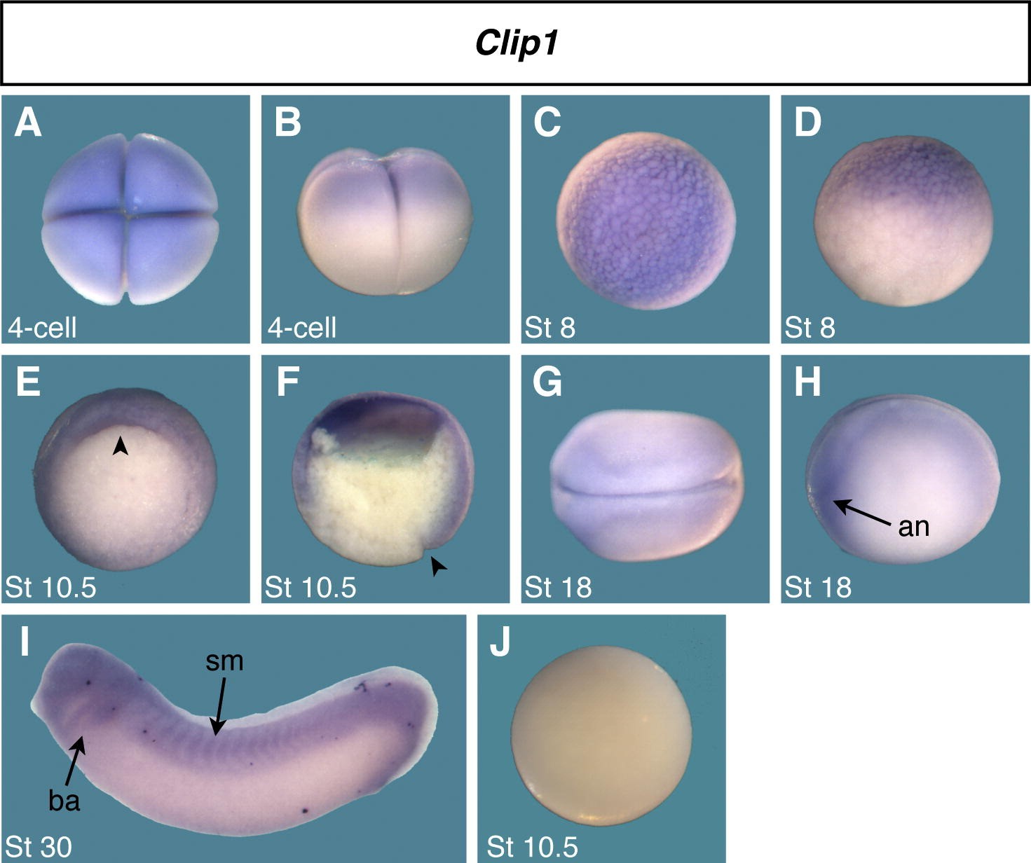
Fig. 3. Spatial expression pattern of Clip1 during early Xenopus development. Maternal Clip1 is localized in animal region and zygotic Clip1 becomes detectable in marginal region from the gastrulation. Clip1 is also expressed in the head region and somite at the tailbud stages. (A and C) Animal view. (B, D, H, and I) Lateral view. (E) Vegetal view. (F) Sagittal bisection view. (G) Dorsal view. (J) Sense control probe. Vegetal view. Arrow heads indicate the dorsal lip of blastopore. an, anterior neural border; ba, branchial arch; sm, somite.

Image published in: Park EC et al. (2012)

Copyright © 2012. Image reproduced with permission of the Publisher, Elsevier B. V.

GeneSynonymsSpeciesStage(s)Tissue
clip1.Lclip-170, clip170X. laevisThroughout NF stage 10.5upper blastopore lip
dorsal marginal zone
blastocoel roof
ectoderm
neuroectoderm
clip1.Lclip-170, clip170X. laevisThroughout NF stage 18neural plate
pre-chordal neural plate border
anterior placodal area
neural tube
neural plate border
clip1.Lclip-170, clip170X. laevisThroughout NF stage 29 and 30somite
pharyngeal arch
mandibular arch
hyoid arch
branchial arch
head region
tail bud
presomitic mesoderm
proctodeum
paraxial mesoderm
clip1.Lclip-170, clip170X. laevisThroughout NF stage 3 (4-cell)animal pole
clip1.Lclip-170, clip170X. laevisThroughout NF stage 8animal hemisphere

Image source: Published

Larger Image
Printer Friendly View

Return to previous page