XB-IMG-124766
Xenbase Image ID: 124766

|
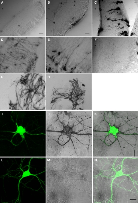
Figure 1. HRP constructs and their expression in cells. Micrographs of vibratome sections through the optic tectum of Xenopus tadpoles in which DAB reactions are used to visualize constructs generated with cDNA from HRP and other proteins as specified: (A) cytosolic HRP. (B) EGFP/ssHRP. (C) EGFP/ssHRPKDEL. (D) EGFP/ssHRP-TM-GFP. (E,F) EGFP/ssHRP-TM. (G) EGFP/ssHRP-XIRbeta-TM. (H) ssHRP-TM-synaptophysin-EGFP. Plasmid definitions are shown in Table 1. (IâN) Expression of mHRP in hippocampal neurons. (IâK) A hippocampal neuron, transfected with EGFP/mHRP. (I) EGFP expression. (J) The mHRP was revealed by DAB reaction. (K) Merge of EGFP and DAB signal. (L,N) A hippocampal neuron, transfected with EGFP only, showed no DAB signal. (L) EGFP expression. (M) Image following DAB reaction. (N) Merge of EGFP and DAB signal. Scale bars in (A) and (B) are 20âμm. Scale bar in (C) is 10âμm and applies to (DâH). Scale bar in (N) is 20âμm and also applies to (IâM). Image published in: Li J et al. (2010) Image downloaded from an Open Access article in PubMed Central. Copyright © 2010 Li, Wang, Chiu and Cline. Larger Image Printer Friendly View |
