XB-IMG-125197
Xenbase Image ID: 125197
|
|
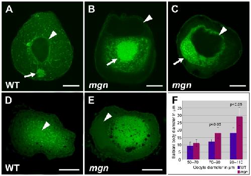
Figure 6. mgn mutant oocytes exhibit an enlarged Balbiani body and absence of mitochondria and ER from the periphery of the oocyte.Oocytes stained with DiOC6 to label ER and mitochondria (A–E). In a wild-type mid stage I oocyte, the nucleus is in the middle of the oocyte (arrowhead) and the Balbiani body is at the future vegetal side of the oocyte (A, arrow). (B,C) Mutant oocytes exhibit an enlarged Balbiani body (arrows), and an absence of ER, mitochondria and the Balbiani body from the periphery. Images are single optical sections. In stage II wild-type oocytes (D), ER and mitochondria are localized throughout the oocyte, whereas in mgn mutant oocytes (E), ER and mitochondria are concentrated in the middle of the oocyte and are absent from peripheral regions. Images are single optical sections of 0.5 micron oocyte sections. (F) Bar graph depicting size of Balbiani body (Bb) during stage I of oogenesis. 50–70 micron oocytes, n = 10 wild-type and 10 mutant oocytes; 70–90 micron oocytes, n = 15 wild-type and 15 mutant oocytes; 90–110 micron oocytes, n = 15 wild-type and 15 mutant oocytes. Arrowheads indicate nuclei. (A–C) scale bars = 25 microns. (D,E) scale bars = 100 microns. Image published in: Gupta T et al. (2010) Gupta et al. Creative Commons Attribution license Larger Image Printer Friendly View |