XB-IMG-117541
Xenbase Image ID: 117541
|
|
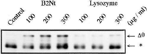
Figure 4. Inhibition of 26S proteasome-catalyzed cyclin B digestion in vitro by the NH2-terminal fragment of Xenopus cyclin B2 (B2Nt). Cyclin Δ0 was incubated with purified 26S proteasome (60 μg/ml) for 120 min in the absence (Control) or presence of various concentrations of B2Nt or lysozyme. Cyclin B was detected with the B63 antibody. The migrating position of the digested cyclin B is indicated by an asterisk. Image published in: Tokumoto T et al. (1997) Image reproduced on Xenbase with permission of the publisher and the copyright holder. Creative Commons Attribution-NonCommercial-ShareAlike license Larger Image Printer Friendly View |