XB-IMG-155000
Xenbase Image ID: 155000
|
|
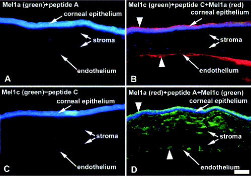
Fig. 3.
Peptide block controls for Mel1a and Mel1c labelling in the Xenopus laevis cornea. (A and C) Corneal sections were incubated with green fluorescent dye conjugated Mel1a or Mel1c receptor antibodies previously pre-incubated with their corresponding peptide and were counterstained with DAPI blue nuclear dye. Absence of green stain indicates complete block of specific antibody binding with specific peptide. (B and D) Corneal sections were incubated with red fluorescent dye-conjugated Mel1a receptor antibody together with green fluorescent dye n conjugated Mel1c receptor antibody previously pre- incubated with either Mel1a or Mel1c peptide and counterstained with DAPI blue nuclear dye. Red labelling indicates areas of specific Mel1a receptor labelling in epithelium and green labelling indicates areas of specific Mel1c receptor labelling in surface epithelium, endothelium (arrowheads) and stroma. Scale bar=100 μm. Image published in: Wiechmann AF and Rada JA (2003) Copyright © 2003. Image reproduced with permission of the Publisher, Elsevier B. V. Larger Image Printer Friendly View |