XB-IMG-125479
Xenbase Image ID: 125479
|
|
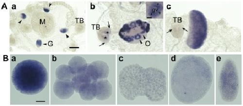
Figure 2. Expression of Clytia Poc1.A. Poc1 mRNA detected by in situ hybridization in adult Clytia medusae. a: Immature female. b: Mature female gonad showing high Poc1 mRNA concentrations in small and medium sized oocytes (inset shows an individual oocyte at higher magnification, with black arrowheads indicating the particulate distribution of the probe around the oocyte nucleus. Scale bar: 50 µm). c: Mature male gonad. Poc1 is expressed strongly in male and female gonads, and weakly at the base of the tentacle bulb (arrows in b and c). G = gonad; M = manubrium; TB = tentacle bulb; O = oocytes; gv = germinal vesicle. Asterisks in b indicate non-specific staining of the tentacle bulb endoderm, frequently observed in Clytia in situs [1]. Scale bar: 100 µm. B. Poc1 mRNA detected by in situ hybridization of eggs, embryos and larvae. Poc1 maternal mRNA is detected strongly in the unfertilized egg (a) but decreases progressively through cleavage (b: 8-cell stage) and blastula (c) stages. The relatively higher signal in gastrula (d) and planula (e) stages probably reflects new trancription starting from the blastula stage. Scale bar: 50 µm. Image published in: Fourrage C et al. (2010) Fourrage et al. Creative Commons Attribution license Larger Image Printer Friendly View |