XB-IMG-198434
Xenbase Image ID: 198434
|
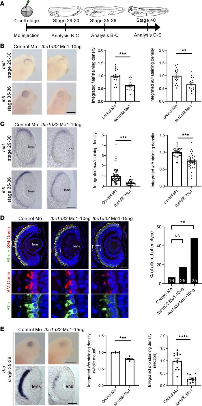
Figure 4. Xenopus RPE and photoreceptor marker expression following tbc1d32 knockdown. (A) Diagram of the experimental design. Whole mount (B) or retinal sections (C) following in situ hybridization against mitf or ihh, respectively, on embryos injected with control Mo or tbc1d32 Mo1. Scatterplots represent the quantification of the integrated density of the staining per eye relative to control Mo; each dot corresponds to 1 eye or 1 section, respectively. (D) Rho and SM opsin immunolabeling on retinal sections of embryos injected with control Mo or embryos injected with 2 doses of tbc1d32 Mo1 (10 or 15 ng). Lower panels, enlargement of the areas indicated by white dashed boxes in the upper panels. The bar plot represents the proportion of eyes with altered staining of Rho and SM opsin for each condition. The number of eyes analyzed per condition is indicated in each bar. Rho, rhodopsin; SM opsin, short and middle wavelength cone opsin. (E) Upper panels, whole-mount in situ hybridization against rhodopsin in embryos injected with control Mo or tbc1d32 Mo1. Lower panels, transverse retinal sections of control and morphant embryos. The scatterplots represent the quantification of the integrated density of rhodopsin staining relative to control Mo; each dot corresponds to 1 eye (left) or 1 section (right). For all scatterplots, data are represented as mean SEM. **P < 0.01; ***P < 0.001; ****P < 0.0001; Fishers exact test (D); 2-tailed Mann-Whitney test (B, C, and E). Scale bars = 400 m for whole-mount embryos and 40 m for sections. Image published in: Bocquet B et al. (2023) © 2023 Bocquet et al. Creative Commons Attribution license
Image source: Published Larger Image Printer Friendly View |