XB-IMG-147102
Xenbase Image ID: 147102
|
|
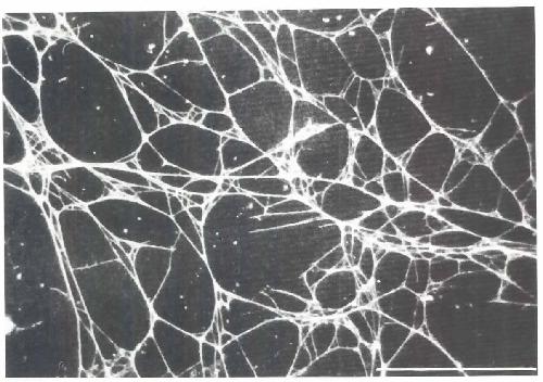
Fig. 3. Fluorescent micrograph of ECM fibrils transferred from the basal suriace ofthe blastocoel roof of an Ambystoma maculatum gastrula to a plastic dish. DeposIted flbnls were stamed with a polyclonal antibody directed against A mexicanum plasma flbronectm, The dorsal lip region of the conditioning explant was on the left and the ammal pole region was on the right Notice that the fibrils are highly aligned parallel to the blastopore-animal pole aXIs (parallel to the long aXIs of the photograph). Migrating mesodermal cells or explanrs of the DMZ adhere to such substrata and migrate preferentially toward the animal pole region of such conditioned areas. Bar: 50 um. Image published in: Boucaut JC et al. (1990) Copyright © 1990. Image reproduced with permission of the Publisher. Larger Image Printer Friendly View |