XB-IMG-124460
Xenbase Image ID: 124460
|
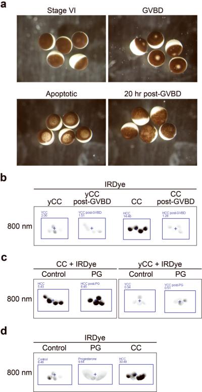
Figure 5. Progesterone-induced oocyte maturation decreases sensitivity to cytochrome c(A) Reflected light images of healthy stage VI oocytes (top left), progesterone-treated oocytes demonstrating GVBD (top right), cytochrome c-injected apoptotic oocytes (bottom left), and deteriorating un-fertilized oocytes 20 hours after GVBD (bottom left). (B) Oocytes were treated with progesterone and allowed to mature until GVBD, approximately 5 hours. Post-GVBD or control-treated oocytes were then injected with the IRDye and yCC or CC (at 67 nM), and then imaged after 1 hour. Average oocyte fluorescence is displayed in Supplemental Figure 1l. (C) Oocytes were treated with progesterone for 30 minutes before injecting them or control-treated oocytes with the IRDye and yCC or CC (at 533 nM), and imaged after 1 hour. Average oocyte fluorescence is displayed in Supplemental Figure 1m. (D) Oocytes were treated with progesterone or control, microinjected with IRDye after GVBD and then imaged for fluorescence 24 hours after progesterone treatment. Oocytes injected at the same time with IRDye and CC at 67 nM were included as a positive control. Average oocyte fluorescence is displayed in Supplemental Figure 1n. Image published in: Johnson CE et al. (2010) Image downloaded from an Open Access article in PubMed Central. Image reproduced on Xenbase with permission of the publisher and the copyright holder. Larger Image Printer Friendly View |