XB-IMG-117755
Xenbase Image ID: 117755

|
|
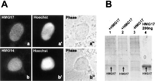
Figure 3. Recombinant HMG-14/-17 proteins accumulate in reconstituted nuclei. (A) Immunofluorescence analysis of reconstituted nuclei after addition of recombinant HMG-17 (a) or HMG-14 (b). Corresponding DNA stain with Hoechst and phase are shown in a′ and b′, and a′′ and b′′, respectively. Bar represents 10 μm. (B) Coomassie stained SDS-gel depicting proteins isolated from purified, reconstituted nuclei (105 nuclei/lane) which were either incubated with (lanes 1 and 2) or without (lane 3) recombinant HMG-17 (arrow). As a marker, 200 ng recombinant HMG-17 protein was loaded in lane 4. The nuclei were incubated in the presence of HMG-17 for either 30 min (lane 1) or 15 min (lane 2). Image published in: Hock R et al. (1998) Image reproduced on Xenbase with permission of the publisher and the copyright holder. Creative Commons Attribution-NonCommercial-ShareAlike license Larger Image Printer Friendly View |
