XB-IMG-126285
Xenbase Image ID: 126285
|
|
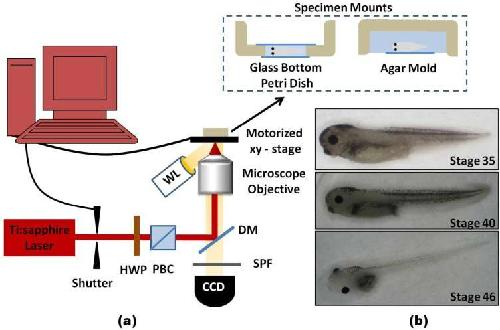
Fig. 1. Schematic of femtosecond laser ablation of Xenopus tadpoles. (a) Femtosecond pulses were focused onto the specimens mounted on top of the motorized stage. For laser ablation, Xenopus laevis younger than stage 40 were held between a Petri dish with a glass welled bottom and a cover slip, while older tadpoles were placed inside a depression made of agar, secured with a glass cover slip, and then inverted for placement on the stage. (HWP—half wave plate, PBC—polarizing beam cube, SPF—short pass filter, DM—dichroic mirror, WL—white light source) (b) Images of Xenopus tadpoles at three different developmental stages. Image published in: Mondia JP et al. (2011) ©2011 Optical Society of America. Creative Commons Attribution-NonCommercial-NoDerivatives license Larger Image Printer Friendly View |