XB-IMG-117352
Xenbase Image ID: 117352
|
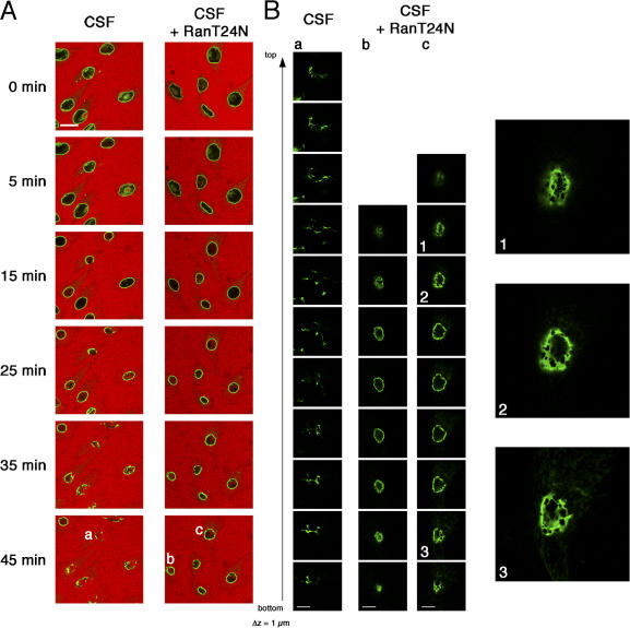
Figure 6. Inhibition of RCC1 activity by RanT24N interferes with final steps of NEBD in vitro. (A) Nuclei of semipermeabilized GFP-LAP2β (green)–expressing HeLa cells were preloaded with either Ran wild-type (left) or RanT24N (right). Then, cells were incubated in CSF extract supplemented with either 20 μM Ran wild-type (left) or RanT24N (right), an energy-regenerating system, and a 155-kD TRITC-labeled dextran (red). NEBD was monitored by time-lapse confocal laser-scanning microscopy. The relative fluorescence intensity inside nuclei (n > 14) was quantified as in Fig. 2. Dextran influx into nuclei of control cells or cells preincubated with RanT24N occurred after 16 min (±3 min) and 19 min (±4 min), respectively. (B) High magnification confocal sectioning of disassembled nuclei from A (indicated by letters) was performed using a 63× objective and a 4× zoom. Z-step width is 1 μm. On the right, representative magnifications of selected frames from the z stacks (indicated by numbers) are shown. Bars (A), 20 μm; (B) 10 μm. Image published in: Mühlhäusser P and Kutay U (2007) Copyright © 2007, The Rockefeller University Press. Creative Commons Attribution-NonCommercial-ShareAlike license Larger Image Printer Friendly View |