XB-IMG-128524
Xenbase Image ID: 128524
|
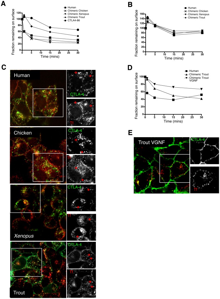
Figure 3. Endocytosis rates of CTLA-4 chimeras.A. CHO cells expressing CTLA-4 chimeras were labeled at 4°C with anti-CTLA-4 to label surface CTLA-4. Cells were then warmed to 37°C to allow endocytosis for the times indicated. Cells were then placed on ice and any remaining surface CTLA-4 detected with Alexa647 anti-mouse IgG. The 647 signal was plotted against time as the fraction remaining compared to 4°C. B. CHO cells expressing CTLA-4 chimeras were labeled as in A but in medium supplemented with sucrose (0.45 M) to prevent endocytosis. C. CHO cells expressing the chimeric CTLA-4 constructs were incubated with a transferrin (Tf) Alexa633 conjugate (Invitrogen) and anti-CTLA-4 PE at 37°C for 45 minutes. Cells were subsequently fixed and analysed by confocal microscopy. The red arrows indicate co-localisation. D. Rate of endocytosis of VGNF mutant was performed as described in A. E. Transferrin uptake of VGNF mutant was performed as described in C and analysed by confocal microscopy. Image published in: Kaur S et al. (2013) Image reproduced on Xenbase with permission of the publisher and the copyright holder. Creative Commons Attribution license Larger Image Printer Friendly View |