XB-IMG-119688
Xenbase Image ID: 119688
|
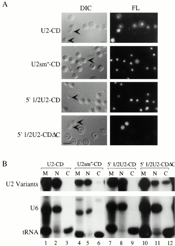
Figure 6. (A) Intranuclear localization of U2-C/D motif chimeras. 1 fmol of fluorescein-labeled wild-type U2-C/D motif chimera (U2+CD), Sm-mutant U2-C/D motif chimera (U2smâ-CD), 5â² 1/2U2-C/D motif chimera (5â² 1/2U2-CD), or 5â² 1/2U2-C/DÎC motif chimera (5â² 1/2U2+CDÎC) was injected into Xenopus oocyte nuclei, and nuclear spreads were prepared 5 h later. DIC and fluorescence (FL) panels are shown for each field. The arrowheads in the DIC panels indicate Cajal bodies. (B) Nucleocytoplasmic distribution of the U2-C/D motif chimeras. The distribution of the injected RNAs within the nuclear and cytoplasmic oocyte compartments was determined as described in the legend to Fig. 4 B. Bar, 10 μm. Image published in: Yu YT et al. (2001) © 2001 The Rockefeller University Press. Creative Commons Attribution-NonCommercial-ShareAlike license Larger Image Printer Friendly View |