XB-IMG-128753
Xenbase Image ID: 128753
|
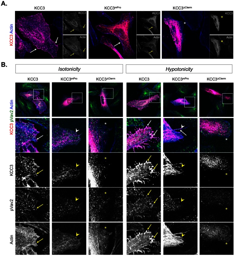
Figure 5. KCC3 colocalizes with the active form of Vav2 in actin-rich membrane protrusions induced by hypotonic conditions.(A) Distribution of wild-type and mutant KCC3 forms in transiently transfected HeLa cells. Note the aberrant distribution of the KCC3 mutant forms in the cytoplasm. (B) The active form of Vav2 accumulates with wild-type KCC3 but not with KCC3mPro or KCC3ΔCterm at the cell periphery. Wild-type KCC3 accumulates with pVav2 in actin-rich plasma membrane protrusion under hypotonic conditions (left panel). These results were obtained after a 10 min treatment either in an isotonic or in a hypotonic medium. In the merged images, KCC3 reactivity is indicated in red, pY-Vav2 is indicated in green and polymerized actin detected by phalloidin is indicated in blue. Arrows indicate co-localisation; the stars (*) indicate actin-rich membrane not showing KCC3 immuno-detection; the arrowheads indicate actin-rich membrane showing KCC3 immuno-reactivity but not pVav2 co-localisation. Image published in: Salin-Cantegrel A et al. (2013) Image reproduced on Xenbase with permission of the publisher and the copyright holder. Creative Commons Attribution license Larger Image Printer Friendly View |