XB-IMG-122355
Xenbase Image ID: 122355
|
|
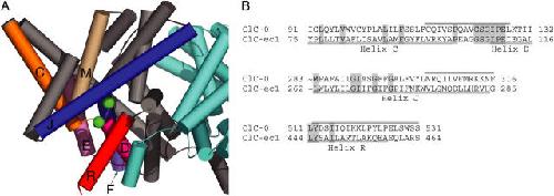
Figure 1. . Candidate pore-lining secondary structures. (A) Structure of ClC-ec1 (1OTS) as viewed from within the membrane. The subunits are colored gray and cyan, with secondary structures lining the intracellular vestibule of the gray subunit colored as follows: helix C and loop CD, orange; helix D and loop DE, magenta; helix E, light pink; helix F, periwinkle; helix J, royal blue; helix M, tan; helix R, red. The figure was created using Pymol (DeLano, 2002). (B) Alignment of secondary structures screened. ClC-ec1 and ClC-0 sequences were aligned using Clustal-W, with slight manual adjustment. Conserved residues are shaded. The regions screened are indicated by bars above the ClC-0 sequence. The beginning of helix J is omitted. Image published in: Engh AM and Maduke M (2005) Copyright © 2005, The Rockefeller University Press. Creative Commons Attribution-NonCommercial-ShareAlike license Larger Image Printer Friendly View |