XB-IMG-117673
Xenbase Image ID: 117673
|
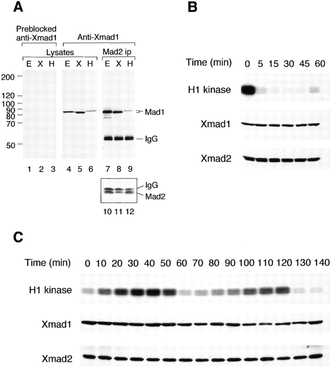
Figure 6. Xmad1 abundance is constant throughout the cell cycle. (A) Specificity of anti-Xmad1 antibodies in immunoblots of frog egg extracts (lanes E), frog cultured cells XTC (lanes X), and HeLa cells (lanes H). An affinity-purified anti-Xmad1 antibody (lanes 4–9) or the same antibody preblocked with recombinant Xmad1 protein (lanes 1–3) were used. Lanes 1–6, egg extracts or cell lysates. Lanes 7–12, anti-Xmad2 immunoprecipitates prepared from egg extracts or cell lysates were immunoblotted with anti-Xmad1 (lanes 7–9) or anti-Xmad2 (lanes 10–12) antibodies. (B) CSF-arrested extract (time 0) was incubated with calcium for the time indicated on top, and immunoblotted for Xmad1 (middle) or Xmad2 (bottom). Top, histone H1 kinase assay. (C) Immunoblots of cycling extracts using antibodies specific to Xmad1 (middle) or Xmad2 (bottom). Mitosis occurs at ∼50 and 120 min as determined by histone H1 kinase assay (top) and by the nuclear morphology (data not shown). Image published in: Chen RH et al. (1998) Image reproduced on Xenbase with permission of the publisher and the copyright holder. Creative Commons Attribution-NonCommercial-ShareAlike license Larger Image Printer Friendly View |