XB-IMG-159719
Xenbase Image ID: 159719
|
|
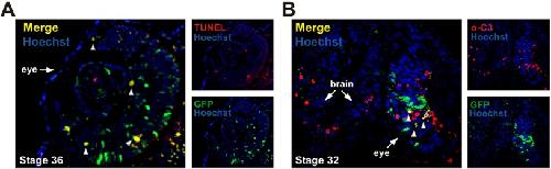
Fig. 4. DEVD reporter activity colocalizes with TUNEL and active casp3 staining. Confocal images of cryosectioned eyes isolated at stages (A) 36 and
(B) 32, when massive cell death is occurring in the developing retina (X. tropicalis). Arrowheads indicate GFP-positive retinal cells colocalizing with TUNEL-positive
cells (A) or active casp3 (B) during retinogenesis. Arrows indicate the eye and the brain on the sections. Image published in: Tran HT et al. (2017) Copyright © 2017. Image reproduced with permission of the Publisher, The Company of Biologists Ltd. Larger Image Printer Friendly View |