XB-IMG-122727
Xenbase Image ID: 122727

|
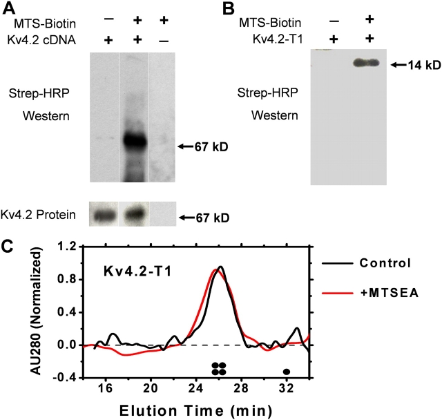
Figure 8. Biochemical evidence of the chemical modification of Kv4.2 and Kv4.2-T1 by MTSEA-biotin. (A) Membrane fragments containing Kv4.2 and KChIP3 (Kv4.2:KChIP3, 1:3) were reacted with MTSEA-biotin for 20 min at room temperature, and then electrophoresed and blotted with either anti-Kv4.2 or streptavidin-HRP (MATERIALS AND METHODS). (B) Likewise, the purified T1 domain of Kv4.2 was also reacted with the biotinylated MTSEA reagent and screened with streptavidin-HRP. The indicated molecular weights correspond to those of Kv4.2 a-monomer (67 kD) and the monomeric Kv4.2-T1 protein (14 kD). (C) FPLC profile of the Kv4.2-T1 protein before (black) and after (red) treatment with MTSEA-biotin (0.5 mM). AU280, normalized absorbance units at 280 nM. The expected elution times of the T1 tetramer and the T1 monomer are schematically marked above the abscissa. Image published in: Wang G et al. (2005) Copyright © 2005, The Rockefeller University Press. Creative Commons Attribution-NonCommercial-ShareAlike license Larger Image Printer Friendly View |
