XB-IMG-126950
Xenbase Image ID: 126950
|
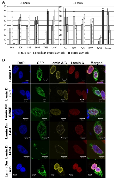
Figure 6. Farnesylation incompetent lamin Dm mutants do not localize efficiently to nuclear lamina and nuclear envelope.Localization of fusion GFP-lamin Dm and mutant proteins after 24 h (A) and 48 h (B) post-transfection into HeLa cells visualized under a confocal microscope and quantitative analyses of appearance of the particular phenotypes. Cells were stained for DNA with DAPI, for endogenous lamin A/C with mouse monoclonal antibodies Jol-2 and for lamin C alone with rabbit affinity purified antibodies. Staining with secondary antibodies was with goat anti-mouse secondary antibodies conjugated with TRITC and goat anti-rabbit secondary antibodies conjugated with Cy-5 respectively. Lamin Dm S25E and T435E were visualized by eGFP fluorescence. Single confocal Z-sections are shown through the center of nuclei. All typically observed phenotypes are shown for lamin Dm T435E mutant. Note the localization of lamin Dm T435E in the cytoplasm only. Image published in: Zaremba-Czogalla M et al. (2012) Zaremba-Czogalla et al. Creative Commons Attribution license Larger Image Printer Friendly View |