XB-IMG-129695
Xenbase Image ID: 129695

|
|
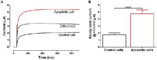
Figure 2. Voltage-gated ion channel activated during apoptosis.A: Electrophysiological properties of an outward current in control oocytes (black) and staurosporine (1 µM) treated oocytes (red) in Xenopus oocytes at +100 mV. The difference between the current in control and STS-treated oocytes is also plotted (dashed line). B. Mean steady-state current at +100 mV in control (1.8 µA±0.5 µA, n = 17) and apoptotic oocytes treated with 1 µM STS (4.8 µA±0.3 µA, n = 13). Steady state currents at +100 mV are significantly larger in the apoptotic oocytes than in controls (**** P<0.0001). Statistical analyses are mean ± SEM and unpaired t-test. Recordings were done in 100Na solution. Image published in: Englund UH et al. (2014) Image reproduced on Xenbase with permission of the publisher and the copyright holder. Creative Commons Attribution license Larger Image Printer Friendly View |
