XB-IMG-125940
Xenbase Image ID: 125940
|
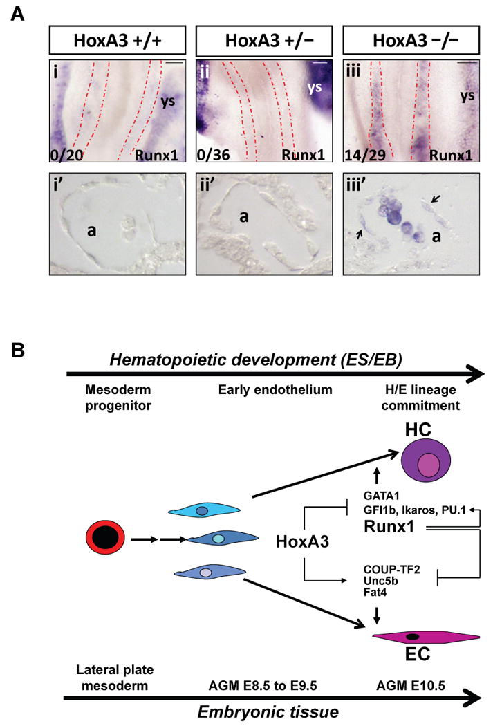
Figure 5. Regulation of Runx1 by HoxA3(A) In situ hybridization showing Runx1 expression in HoxA3 +/+, +/− and −/− E8.5 embryos. Runx1 expression is absent in the dorsal aortae of HoxA3 +/+ (i) and HoxA3+/− (ii) embryos, but robustly expressed in yolk sac. Runx1 is ectopically expressed in the dorsal aortae of HoxA3−/− (iii) embryos. Stippled red lines outline dorsal aortae. Penetrance of this phenotype is indicated at lower left. (i′-iii′) Sections of the embryos shown above. Both endothelial and hematopoietic cells are negative for Runx1 in wild-type or heterozygous embryos, while Runx1-expressing cells are found in HoxA3−/− embryos. Arrows indicate Runx1-expressing endothelial cells. a, aorta; ys, yolk sac. Scale bar = 50 μm for whole mounts, 10 μm for sections. (B) Model for regulation of endothelial hemogenesis by HoxA3 and Runx1. HoxA3 represses a cascade of transcription factors that promote hemogenesis and induces a set of genes that maintain endothelial character. Runx1 is a positive regulator of most of these transcription factors, and a negative regulator of genes essential for endothelial character, thus transient expression of Runx1 erases the endothelial program and initiates the hematopoietic. Image published in: Iacovino M et al. (2011) Image downloaded from an Open Access article in PubMed Central. Image reproduced on Xenbase with permission of the publisher and the copyright holder. Larger Image Printer Friendly View |