XB-IMG-123851
Xenbase Image ID: 123851
|
|
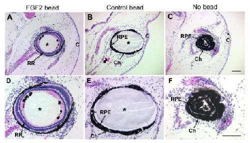
Figure 4. The RPE is a likely source of retinal regeneration. The anterior third of the eye was dissected out, and the neural retina was removed from the posterior eyecup of Xenopus laevis tadpoles, at which point either an FGF-2-soaked bead (A, D), a control bead (B, E), or no bead (C, F) was introduced in eyecups. The panel shows histological sections of eyes collected 30 days postsurgery and stained with hematoxylin and eosin. D-F are higher magnification images of A-C respectively. Robust retinal regeneration was observed in all eyes treated with FGF-2 (A, D), whereas there was no retinal regeneration in any case of eyes exposed to control beads (B, E) or no bead at all (C, F). Abbreviations: choroid layer (Ch); retinal pigmented epithelium (RPE); regenerated retina (RR), cornea (C). Asterisks indicate beads. Scale bars represent 100 μm (scale bar in C applies to A-C and scale bar in F applies to D-F). Image published in: Vergara MN and Del Rio-Tsonis K (2009) Image reproduced on Xenbase with permission of the publisher and the copyright holder. Creative Commons Attribution license Larger Image Printer Friendly View |