XB-IMG-127196
Xenbase Image ID: 127196
|
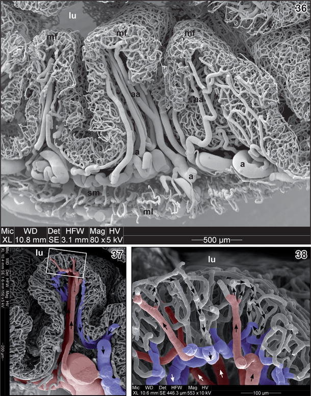
Figs. 36â38. Fig. 36 Microvascular pattern of gastric mucosal folds (mf) in adult Xenopus. Detail from a transverse section. Note strongly undulating arteries (a) with ascending arterioles (aa). lu Lumen, ml muscular layer (muscularis), sm submucosa. Fig. 37 Microvascular pattern of a single mucosal fold. Detail from Fig. 33 (boxed area). Note two ascending arterioles (colored red) and two descending venules (colored blue) supplying/draining the mucosal capillary bed. Arrows indicate direction of blood flow. lu Lumen. Fig. 38 Microvascular pattern of the mucosa of the stomach in adult Xenopus. Detail from Fig. 37 (boxed area). Note that ascending arterioles (colored red) capillarize close below the mucosal epithelium. Mucosal capillaries descend centrifugally and drain into venules (colored blue) located at the base of the mucosa. Arrows indicate direction of blood flow. lu Lumen Image published in: Lametschwandtner A et al. (2012) © The Author(s) 2012. Creative Commons Attribution-NonCommercial license Larger Image Printer Friendly View |