XB-IMG-128996
Xenbase Image ID: 128996

|
|
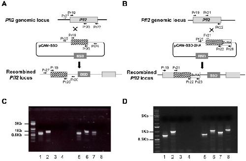
Figure 3. Targeted gene disruption and HA-tagging of the PfI2 locus. A. Disruption of PfI2 by knock-out strategy using single homologous recombination. The pCAM-BSD construct, the blasticidin-resistance cassette (BSD), the location of the primers (Additional file 1: Table S1) used for PCR analysis and the locus resulting from integration are indicated. B. Insertion of an HA epitope tag at the PfI2 C terminus (Knock-in strategy). C. Analysis of pCAM-BSD-PfI2 transfected 3D7 culture by PCR (Knock-out strategy). Lanes 1–4 correspond to DNA extracted from wild type parasites, lanes 5–8 to DNA extracted from transfected parasites. Lanes 1 and 5 represent the detection of a portion of wild type locus (Pr19 and Pr20); lanes 2 and 6, the detection of wild type locus (Pr19 and Pr22); lanes 3 and 7 show the detection of episomal DNA (Pr25 and Pr26) and lanes 4 and 8 show the detection of the integration at the 5′end of the insert (Pr27 and Pr26). The absence of PCR product amplification using genomic DNA prepared from transfected parasite culture (Pr27 and Pr26) indicated the lack of homologous recombination (lane 8). D. PCR analysis of pCAM-PfI2-2HA transfected 3D7 culture (Knock-in strategy)). Lanes 1–4: DNA extracted from wild type parasites; lanes 5–8: DNA extracted from transfected parasites. Lanes 1 and 5: detection of a portion of wild type locus (Pr21 and Pr22); lanes 2 and 6: detection of wild type locus (Pr19 and Pr22); lanes 3 and 7: detection of episomal DNA (Pr25 and Pr28) and lanes 4 and 8: detection of the integration at the 3′end of the insert (Pr19 and Pr28). The amplification of a PCR product at ~ 1000 pb using genomic DNA prepared from transfected parasite culture (Pr19 and Pr28) indicated the homologous recombination and integration of the 2-HA construct in endogenous PfI2 (lane 8). Image published in: Fréville A et al. (2013) Copyright ©2013 Frèville et al. Creative Commons Attribution license Larger Image Printer Friendly View |
