XB-IMG-125482
Xenbase Image ID: 125482
|
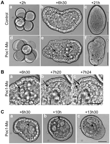
Figure 5. Poc1 MO disrupts cell division.Images from time-lapse recordings of Clytia embryos injected with Poc1-MO before fertilization (Movie S1). Times post fertilization are indicated. A. Cleavage divisions in Poc1-MO embryos proceeded in line with uninjected controls (a,d). Starting from around 6 hours post fertilization (blastula stage: b,e), Poc1-MO embryos showed lengthening of the cell cycle in some cells, followed by cell division defects/arrest, resulting in the appearance of larger cells (arrowheads). The accumulation of large cells in the blastocoel contributed to disruption and delay of gastrulation, resulting in characteristically deformed planula larvae (c,f). B. Cell cycle lengthening in a Poc1-MO blastula. Arrows identify a cell already larger than those around it cell which remains un-divided for at least 50 minutes (between a and b) before dividing (c). In uninjected controls filmed in parallel, cell division was largely synchronous until the blastula stage, with division occuring every 30 minutes (Movie S1, Figure 5A). C. Accumulation of abnormally large cells (arrowheads) in the blastocoel cavity (a), which remained undivided during gastrulation (b, c). Scale bars: 100 µm. Times indicated are hours post fertilization at 18°C. Image published in: Fourrage C et al. (2010) Fourrage et al. Creative Commons Attribution license Larger Image Printer Friendly View |