XB-IMG-125951
Xenbase Image ID: 125951
|
|
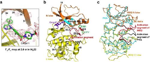
Figure 2. Inhibitor bound xIKKβ kinase domain (KD)a, Fo-Fc electron density map for Cmpd1 in the I4122 structure, contoured at 2.0 σ. Carbon, nitrogen and oxygen atoms are shown in green, blue and red, respectively. b, Structure of xIKKβ KD. Glycine-rich loop: cyan; activation segment: red except that the DLG and APE motifs are in black; Cmpd1: purple. Side chains of phosphomimic residues E177 and E181 are shown. c, Superposition between xIKKβ (orange and yellow) and PKA (cyan, PDB code 1ATP). The activation segments of xIKKβ and PKA are shown in red and black, respectively. Image published in: Xu G et al. (2011) Image downloaded from an Open Access article in PubMed Central. Image reproduced on Xenbase with permission of the publisher and the copyright holder. Larger Image Printer Friendly View |