XB-IMG-128714
Xenbase Image ID: 128714
|
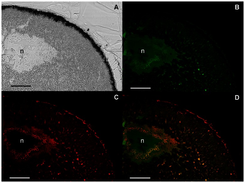
Figure 3. Polarization of GIRK5 within the animal pole.A) Light transmission image of the oocyte animal pole section; B,C,D) Confocal microscopic image of the same animal pole section visualizing EGFP-GIRK5 (B, green), ECFP-ER (C, red) or both (D). GIRK5 localized both in the nucleus (n, green) and the ER (yellow). A 40× objective was used. Scale bar: 100 µm. Image published in: Díaz-Bello B et al. (2013) Image reproduced on Xenbase with permission of the publisher and the copyright holder. Creative Commons Attribution license Larger Image Printer Friendly View |