XB-IMG-120095
Xenbase Image ID: 120095

|
|
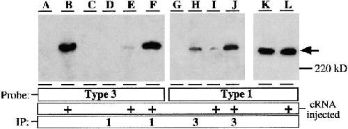
Figure 1. Expression of endogenous type 1 and recombinant type 3 InsP3 receptors in Xenopus oocytes. (A–F) Immunoblotted with Ab-3; (G–K) immunoblotted with Ab-1. (A) Western blot of lysate from an equivalent of one control oocyte, demonstrating lack of endogenous type 3 InsP3R expression; (B) Western blot of lysate from an equivalent of one r-InsP3R-3 cRNA-injected oocyte, demonstrating expression of the recombinant r-InsP3R-3 protein; (C) Western blot of type 3 InsP3R adsorbed nonspecifically to agarose beads, from lysate from fifteen control oocytes; (D) Western blot of type 3 InsP3R in InsP3R-1 immunoprecipitate, from lysate from fifteen control oocytes; (E) Western blot of type 3 InsP3R adsorbed nonspecifically to agarose beads, from lysate from fifteen cRNA-injected oocytes; (F) Western blot of type 3 InsP3R in InsP3R-1 immunoprecipitate from lysate, from fifteen cRNA-injected oocytes. Difference between F and E represents the amount of recombinant r-InsP3R-3 specifically immunoprecipitated with X-InsP3R-1. (G) Western blot of X-InsP3R-1 adsorbed nonspecifically to agarose beads, from lysate from fifteen control oocytes; (H) Western blot of X-InsP3R-1 in InsP3R-3 immunoprecipitate, from lysate from fifteen control oocytes. The InsP3R detected is most likely caused by cross-interaction of the immunoprecipitating Ab-3 with the large amount of endogenous X-InsP3R-1 present in the lysate from fifteen oocytes. (I) Western blot of X-InsP3R-1 adsorbed nonspecifically to agarose beads, from lysate from fifteen cRNA-injected oocytes; (J) Western blot X-InsP3R-1 in InsP3R-3 immunoprecipitate, from lysate from fifteen cRNA-injected oocytes. Difference in protein amounts between H/I and J represents the amount of X-InsP3R-1 specifically immunoprecipitated with recombinant r-InsP3R-3. (K) Western blot of X-InsP3R-1 in lysate from an equivalent of one control oocyte; (L) Western blot of X-InsP3R-1 in lysate from an equivalent of one cRNA-injected oocyte. Comparisons of K and L indicate that r-InsP3R-3 expression does not induce changes in the expression levels of the endogenous X-InsP3R-1. Western blots of type 1 InsP3R in InsP3R-1 immunoprecipitates or type 3 InsP3R in InsP3R-3 immunoprecipitates are not shown because too much protein was present in those lanes for the protein bands to be properly detected with the same exposure as the other lanes. Image published in: Mak DO et al. (2000) © 2000 The Rockefeller University Press. Creative Commons Attribution-NonCommercial-ShareAlike license Larger Image Printer Friendly View |
