XB-IMG-126041
Xenbase Image ID: 126041
|
|
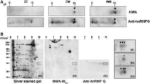
Figure 5. Interaction of the P. waltl hnRNP G isoforms with the WEc RNA. (A) 2D-NWA of the 32P-labelled RNA WEc probe with nuclear proteins from ZZ, ZW and WW GVs, followed by immunodetection using the anti-hnRNP G serum. Note that the homologues of hnRNPG formed a train of basic spots ranging from pI 9 to 10 with two major spots at pI 10. The most basic spot corresponded to the one detected by NWA in ZW and WW GVs. (B) An example of isoelectrofocusing of the protein extracts of the ZW GVs showing three spots of hnRNP G at pI 10 (arrowheads) in the silver stained gel (S). Note that only one spot was visible in the northwestern blot (N). Image published in: Kanhoush R et al. (2011) © The Author(s) 2011. Creative Commons Attribution-NonCommercial license Larger Image Printer Friendly View |