XB-IMG-125338
Xenbase Image ID: 125338
|
|
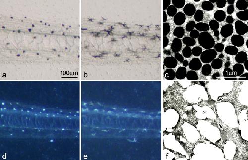
Fig. 1. Pigment cells present in the wild-type tadpole tail (aâc) and the mutant tadpole tail (dâf) at stage 48. a The wild-type tail placed in BSS before α-MSH administration (transmitted light). b The wild-type tail after α-MSH administration (1 μg/ml) (transmitted light). c Ultrastructure of melanophores of the wild-type. d The mutant tail placed in BSS before α-MSH administration (incident light). e The mutant tail after α-MSH administration (1 μg/ml) (incident light). f Ultrastructure of white pigment cells in the mutant. While wild-type melanophores were filled with many mature melanosomes (c), white pigment cells in the mutant contained both irregular reflecting platelets (f, asterisk) and premelanosomes with internal lamellar structures (f, arrows). Note that white pigment cells (d,e) in the mutant responded to α-MSH and dispersed pigment organelles in the same manner as wild-type melanophores (a b) Image published in: Fukuzawa T (2010) © The Author(s) 2010. Creative Commons Attribution-NonCommercial license Larger Image Printer Friendly View |