XB-IMG-119687
Xenbase Image ID: 119687
|
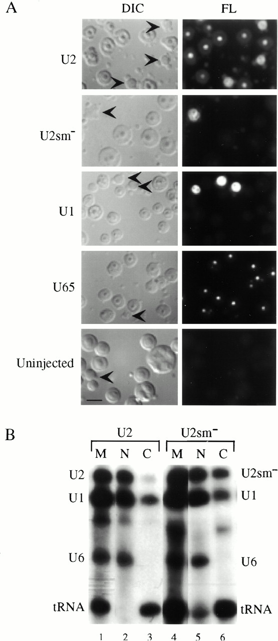
Figure 4. (A) Intranuclear localization of wild-type U2 and Sm-mutant U2 (U2smâ) snRNAs. 1 fmol of 32P- and fluorescently labeled, in vitroâtranscribed U2, Sm-mutant U2, U1, or U65 was injected into Xenopus oocyte nuclei. Nuclear spreads were prepared 5 h later. U1 was included as a positive control for Cajal body localization and as a negative control for nucleolar localization (Narayanan et al. 1999). U65 snoRNA served as a positive control for nucleolar localization and as a negative control for Cajal body localization (Narayanan et al. 1999). A nuclear spread prepared from an uninjected oocyte was included to control for background fluorescence of the preparations. The nuclear spreads were analyzed by DIC and fluorescence (FL) microscopy. Each panel includes several nucleoli and a few Cajal bodies. Cajal bodies are indicated by arrowheads in the DIC panels. (B) Nucleocytoplasmic distribution of U2 and Sm-mutant U2 (U2smâ) snRNAs. Injected oocytes from the same batch analyzed above were dissected into nuclear and cytoplasmic fractions after 5 h. Samples were analyzed by denaturing PAGE and autoradiography to determine the stability and nucleocytoplasmic distribution of the injected RNAs. tRNA was used as a positive control for export and U6 served as a nuclear retention control (Narayanan et al. 1999; Speckmann et al. 1999). Nuclear (N) RNAs are in lanes 2 and 5, cytoplasmic (C) RNAs are in lanes 3 and 6, and marker (M) lanes 1 and 4 show RNAs before injection. Bar, 10 μm. Image published in: Yu YT et al. (2001) © 2001 The Rockefeller University Press. Creative Commons Attribution-NonCommercial-ShareAlike license Larger Image Printer Friendly View |