XB-IMG-125006
Xenbase Image ID: 125006

|
|
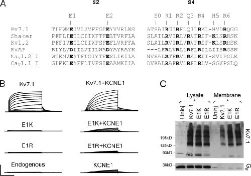
Figure 1. Sequence alignment of S2 and S4 from various voltage-dependent ion channels and proteins. (A) Conserved negatively charged residues in S2 and positively charged residues in S4 are in bold. (B) Currents generated from WT, E1K, and endogenous channels. Oocytes were held at −80 mV, depolarized from −80 to +60 mV for 5 s, and repolarized at −40 mV for 3 s. Scale, 4 µA for all except Kv7.1+KCNE1 (20 µA); 2 s for all currents in this and subsequent figures. (C) Western blot probing for Kv7.1 and Gβ in the whole cell lysate and biotinlyated membrane fraction from oocytes. Gβ is a cytoplasmic protein. Black lines indicate that intervening lanes have been spliced out. Image published in: Wu D et al. (2010) © 2010 Wu et al. Creative Commons Attribution-NonCommercial-ShareAlike license Larger Image Printer Friendly View |
