XB-IMG-124401
Xenbase Image ID: 124401

|
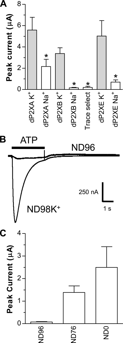
FIGURE 1. dP2X receptors are inhibited by Na+. Two-electrode voltage clamp recordings at a holding membrane potential of −70 mV were made from Xenopus oocytes expressing dP2X receptors. A, comparison of peak current amplitudes recorded in response to 3 mm ATP in extracellular recording solutions consisting either of ND96 (Na+ based) or ND98K+ (K+ based) with both solutions at pH 6.2. Note inhibition of currents by Na+ particularly for dP2XB and dP2XE channels. “Trace select” denotes ND96 extracellular recording solution made with ultrapure NaCl (Fluka 38979) for dP2XB. *, significant difference, p < 0.05, compared with respective ND98K+ current (n = 7). B, direct comparison of current amplitudes in ND96 and ND98K+ extracellular recording solutions (both pH 6.2) in the same dP2XB-expressing oocyte. Application of ATP (3 mm indicated by the bar) in ND96 was followed 5 min later by ATP application in ND98K+. C, mean current amplitudes in dP2XB-expressing oocytes after substitution of Na+ in ND96 with NH4+. ND76 denotes ND96 with 20 mm NH4Cl substituting for 20 mm NaCl. ND0 denotes solution with zero Na+ and 96 mm NH4Cl (n = 7). Image published in: Ludlow MJ et al. (2009) © 2009 by The American Society for Biochemistry and Molecular Biology, Inc. Creative Commons Attribution-NonCommercial license Larger Image Printer Friendly View |
