XB-IMG-122555
Xenbase Image ID: 122555
|
|||||||||||||||||||||||||||||||||||
|
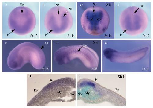
Figure 1. Expression of skp2 and Xic1. Whole-mount in situ hybridisation at indicated stages show expression of (a,b,d,e,g) skp2 and (c,f) Xic1. (a,c) Dorsal view with anterior toward the bottom. (b,d) Anterior view with dorsal toward the top. (e-g) Lateral view with anterior to the left. (h,i) Expression of skp2 and Xic1, respectively, in a vibratome section of stage 16 embryos; primary neurons are indicated with black arrows. Ep, epidermis; I, intermediate stripe; L, lateral stripe; M, medial stripe; My, myotome; Nf, neural folds; Not, notochord; Np, neural plate; Nt, neural tube; P, placodes. Image published in: Boix-Perales H et al. (2007) Copyright © 2007 Boix-Perales et al. Creative Commons Attribution license
Image source: Published Larger Image Printer Friendly View |