XB-IMG-130814
Xenbase Image ID: 130814
|
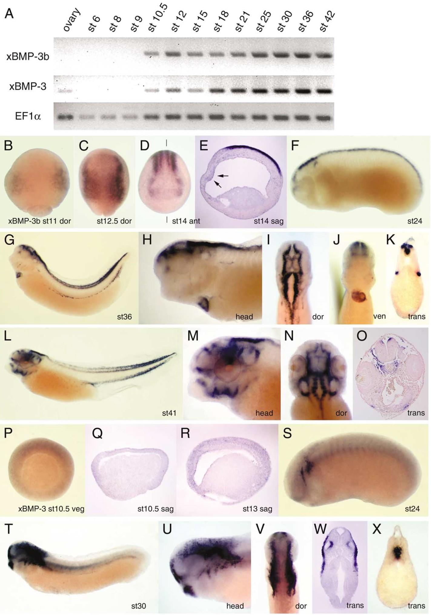
Fig. 2. Xenopus BMP-3b and BMP-3 are expressed in dorsal ectodermal and mesodermal tissues. (A) Temporal expression analyzed by RT-PCR. BMP-3b
and BMP-3 are expressed at quite low levels. After whole-mount in situ hybridization, embryos were stained for 6 days to detect xBMP-3b (BâO) and
overnight for xBMP-3 (PâX). (B) Dorsal view of stage 11 embryo. Anterior region faces top. BMP-3b expression appears on dorsolateral ectoderm before
neural plate is visible. Expression extends to entire neural plate until stage 12.5 (C), and localizes upon four stripes at neural plate midline and edge. (D)
Expression in anterior neural plate at stage 14. Solid line indicates level of section. (E) Sagittal section of embryo. Anterior faces left. BMP-3b expression
is intense in ectoderm and faint in mesoderm and endoderm. Arrows highlight BMP-3b expression in prechordal plate. (F) Lateral view of stage 24 embryo.
BMP-3b is expressed in hindbrain and dorsal side of otic vesicle and neural tube. (G) BMP-3b is expressed in lips of somites and transiently in the heart.
(H) Close-up view of (G). (I) Dorsal view of (H). (J) Ventral view of (H) highlights BMP-3b expression in ventricular chamber. (K) Transverse section
through trunk of embryo in (H). BMP-3b is expressed in both epaxial and hypaxial dermamyotome, and dorsal half of neural tube. (L) At stage 41, expression
in dermamyotome is restricted to tail. (M) Close-up of head shown in (L). BMP-3b is expressed in dorsal side of otic vesicles, optic cup, nasal pit, stomodeum,
and ventral visceral pouches. (N) Dorsal view of embryo shown in (M). (O) Transverse section of embryo shown in (L). BMP-3b is expressed in head
mesoderm around notochord and neural tube. (P) Vegetal view of stage 10.5 embryo. Dorsal faces top. BMP-3 is expressed in ectoderm and mesoderm. (Q)
Sagittal section of embryo shown in (P). Dorsal side is to right. (R) Sagittal section of stage 15 embryo. Anterior faces left. BMP-3 is expressed in prechordal
plate and chordamesoderm. (S) BMP-3 expression appears around otic vesicle at stage 24. (T) At stage 30, BMP-3 is expressed in cranial neural crest
derivatives and cement gland. (U) Close up view of head shown in (T). (V) Dorsal view of (U). (W) Transverse section through head in (T). BMP-3 is
localized in anterior neurocranium and dorsomedial wall of otic vesicle. (X) Transverse section through trunk. BMP-3 expression is restricted to notochord. Image published in: Hino J et al. (2003) Copyright © 2003. Image reproduced with permission of the Publisher, Elsevier B. V.
Image source: Published Larger Image Printer Friendly View |