XB-IMG-134762
Xenbase Image ID: 134762
|
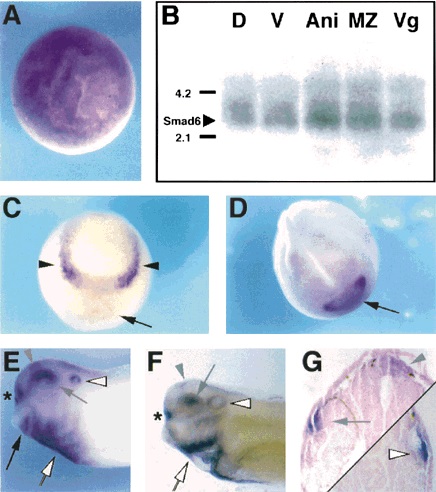
Figure 2. The spatial pattern of expression of Smad6 in developing Xenopus embryos analysed by whole-mount in situ and Northern blot hybridizations. (A) Animal-pole view of a gastrula stage (stage 10) embryo showing uniform Smad6 expression. (B) 10 μg of RNA dissected from the dorsal (D) or ventral (V) halves, or from the animal (Ani), marginal zone (MZ) or vegetal (Vg) thirds of early (stage 10) gastrulae was subjected to Northern blot analysis to detect Smad6 transcripts. The position of 18S (2.1 kb) and 28S (4.2 kb) ribosomal RNAs is indicated to the left of the blot. (C) Anterior-dorsal view of a neurula stage (stage 15) embryo showing Smad6 expression in lateral regions of the anterior neural plate (arrowheads) and weak expression in the prospective cement gland region (arrow) which becomes much stronger by stages 17â18 (D; anterior-dorsal view) and is restricted to the ventral half of the cement gland by stage 30 (E; lateral view, black arrow). (F) Lateral view of a stage 36 embryo cleared in benzyl benzoate:benzyl alcohol (2:1). (G) Transverse sections of stage 36 embryos at the levels of the eye (left) and otic vesicle (right). (EâG) Staining of the dorsal neural tube (grey arrowheads); dorsal eye (grey arrows); lateral otic vesicle (white arrowheads); olfactory placodes (asterisks) and heart anlage (white arrow) is denoted. Image published in: Nakayama T et al. (1998) Copyright © 1998. Image reproduced with permission of the Publisher, John Wiley & Sons.
Image source: Published Larger Image Printer Friendly View |